Immunology Assignment: The Role Of Immunosenescence In Aging Concerning T Cell Responses In Elderly
Question
Task:
Research project: In this immunology assignment, you are required to write a scientific article individually that would be similar to a brief report publication in a peer reviewed scientific journal. You will use the data generated from the flow cytometry and/or ELISA practical sessions. The main issues for this report are data analysis, interpretation and presentation of the findings in a scientific format.
Answer
Abstract
Herein immunology assignment, in the introduction chapter, a brief background of the research area has been illustrated with a short rationale and research aim. The current project aims to identify the role of immunosenescence in aging concerning T cell responses in the elder community. Also, in the method section, protocol, reagents, and instruments for ELISA assay have been mentioned. The ELISA assay has been performed by intra-assay reproducibility with symmetrical immune diffusion and intra-cytoplasmic cytokine staining. in the next section, the practical data have been analyzed concerning “Surface-bound platelets” and estimating sensitive thresholds of T cell lines. In this paper mixed-method (both primary quantitative and secondary qualitative) have been chosen. In the discussion and conclusion, part research limitation has been identified along with the possible suggestions of ELISA assay that can assure the accurate outcome.
Introduction
Aging can be characterized by physiological as well as physical frailty and with ongoing age immunity of an individual changes fundamentally. Aging is related to a decline in innate and adaptive immunity. It is observed that autoimmune diseases or cancer used to happen more frequently in the elder community and though several attributes impact this, immunosenescence contributes a major role (Bektas et al. 2017). Elderly individuals are having low-level chronic inflammation as a consequence of continuous exposure towards antigens along with poor immunity increase of pro-inflammatory cytokines production by senescent T cells, macrophages and effector memory cells. Apart from that, aging is related to several changes in immunity parameters, and its response mechanism is also associated with biochemical or anatomical barriers. Immunosenescence can enhance susceptibility to tissue pathology. Accumulation of the senescent cells may develop immune dysfunction, age-related pathologies within the cytolysis T cell compartment. After puberty, the sub-population of phenotypically and functionally memory T cells begin to compile for the response of thymic involution. This sub-population possesses age-related modification in tissues as well as expresses markers (resident-memory T cells) (Eweret al. 2021).
It is evidenced that reduction of T cells in the older population is linked with susceptibility of infection and develop proinflammatory status resulting in activation-induced apoptosis. Homeostasis expression of T cells may dominantly alter the phenotype and genotype of aging humans. It is reported that immunity (particularly T cell Immune system) and CNS interact, crosstalk as well as the impact each other for physiological processes (physiological homeostasis) and disease status in aging. Early immunosenescence can trigger memory loss along with brain aging. Besides, age-linked neuro-degeneration may worsen T cell immunity, upholding the vicious cycle of age-linked multi-system degeneration. Synthesis of autoantibodies has already been proposed as an alternative to thymus involution with decline T cells and an assemblage of clonal T cells with stimulation because of "neoantigens" whilst aging process. Immunosenescence and underlying T cell mechanisms are developing incidents of infectious disease and immune dysfunctions. It is considering one of the major health issues as it can diminish responses, reduce lifespan and decrease immune security among the elderly (Egorov et al. 2018). Influence of T cells over pathogens through life protection to the step-down of T cell pool together with an increase of memory cells with a contractile diverseness of the repertory of T cell receptors (TCR) because of over internal representation of oligoclonal T cell. The research shed light on the response mechanisms of human beings are connected with the immune system along with T lymphocyte (involve in immunity senescence) mediated antigens in aging.
Research question: What is the contribution of immunosenescence in aging in terms of T cell responses?
Aim of the project: This project aims to evaluate the role of immunosenescence in aging concerning T cell responses in the elderly. Moreover, the purpose of the research paper is to identify the impact of age-related resident memory T cells on the central nervous system as well as human-specific tissue pathology. This paper is also going to illustrate the effect of T cells within brain parenchyma in aging over immune cell migration controlled by chemokines in the presence of antigen-activated lymphocytes.
Method
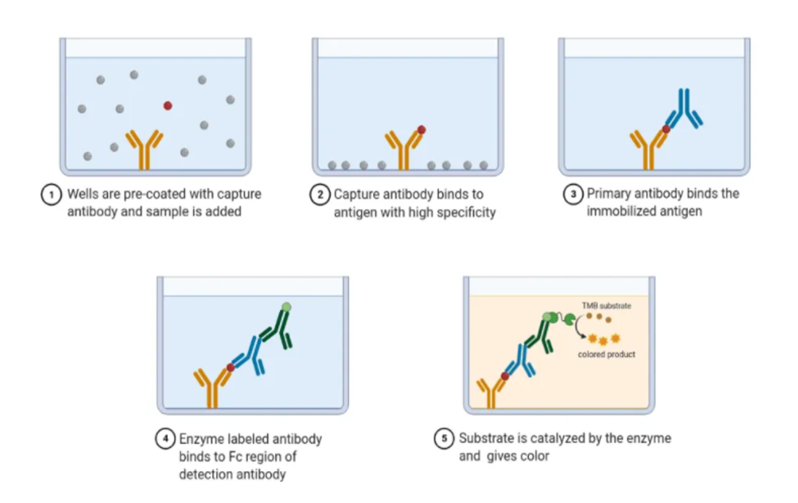
Here, in the research mixed research methods (both primary quantitative and secondary qualitative) have been selected for analyzing practical data gathered from the ELISA method. Also, data have been gathered from medical journals, relevant articles, and books for comparing the practical data. The Enzyme-linked immunosorbent assay (ELISA) deals with rapid tests for quantifying as well as detecting antigens and it involves usage of an enzyme system (Refer to Appendix 1). it is evidenced that the ELISA test is having high sensitivity and specificity as well as it allows the screening of large numbers of samples together. Based on statistical data late differentiated CD8 and CD28 T cells attend in accumulating older people and are responsible for 80% of elderly immunosenescence. CD57 T cell is having senescent, responsible for high-level cytotoxic potential, and acts as a causative attribute for 60% age-related immunosenescence.
Material required
For ELISA test required instruments include pipette, device for washing plates, (semi-automated system) photometer (ELISA reader), and evaluation software. Appropriate filters have been selected as ELISA readers. ELISA reagent kit includes coated plates (to hold immobilize antibody or antigen), control (to standardize the plate), wash concentrates, sample diluent, wash concentrate (buffer solution for washing unbound materials on the plate), substrate, conjugate (react with sample analytes) and stop solution (for color development and finishing enzyme-substrate reaction) (Gurevich et al. 2017). Channel multiprobe has been utilized to transfer peptide or protein within the master plate along with intra-cytoplasmic cytokine staining. T hybridoma cell has been analyzed through flow cytometry, it may provide data regarding cytokine concentration, and T cells have been captured by ELISA solid plate antibody plate.
Flow cytometry
The nuclear factor of activated T cells (NFAT) is involved in producing cytokines and is used for the reporter scheme of T cells activation. This procedure has developed T hybridoma lines which does not decrease by adding fluorescent proteins in a single reaction. T cells are having therapeutic promise and play a significant role in autoimmune disease pathogenesis. Technically through the ELISA proliferation assay, T cell response on peptides have been studied through clinical trial as it allows disaffection of antigen-specific cells. Cytokinin secretion from stimulated T cells can be measured through ELISA assay and alteration of phenotype can be determined through flow cytometry (Ventura MT et al. 2017).
ELISA method
ELISA plate has been washed with wash buffer and then diluting buffer (2% BSA in Phosphate-buffered saline) must be added to it. After that, the effector T cell (TH1 and TH2) has been added and covered with plastic film for incubation (1 hour at 20°C) and accumulation of pathogens. The ELISA method has done through intra-assay reproducibility for IgG, IgE, and IgM with symmetrical immune diffusion. Standard dilution has been prepared with diluted buffer and standard stalk (Gurevich et al. 2017). Flow cytometry has been followed for the quantitative analysis for surface antigen reflection. After getting the cytometry plate it is washed with 150 ?l buffer solution and Alkaline phosphatase-conjugated have been added for incubating for 1 hour. The absorbance has been studied for analyzing the data. Standard co concentration has been followed for the ELISA procedure (1000 ng/ml, 500 ng/ml, 250 1000 ng/ml, and so on). The developed ELISA plate has been scanned followed by the incubation for studying generated T cell lines (in pulsing PBMCs at 107 cells/ml).
Result analysis
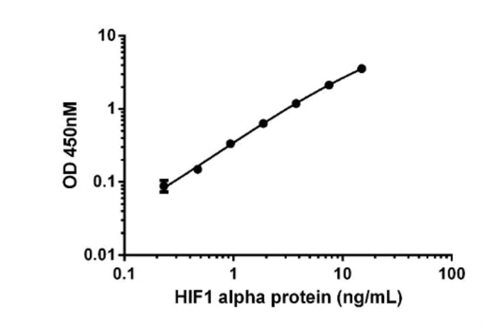
Based on the data from ELISA, T cell responses are limited over cellular standard and in various conditions, the restimulation cycle can show CD4 (cluster of differentiation) as well as CD8 T cell lines (Bektas et al. 2017). For estimating sensitive thresholds, both CD4 and CD8 T cell lines have been diluted into nonresponding PBMCs. Moreover, for demonstrating the usefulness of reference standards, various restimulation cycle T cell lines have been studied. It is observed that the culture supernatant has responded over PBMC stimulation and a standard curve has been developed by using a prism (Refer to appendix 2). Here T cell and the response of neutralizing antibodies (NAb) have been evaluated. Also, T cell responses over asymptomatic infection among the elderly population have been identified along with its direct immunogenic region. Through this practical, “Surface-bound platelets” IgM and IgG have been studied by applying washed platelets and alkaline phosphatase anti-human immunoglobulins. Here the absorbance has been recorded at 405 nm wavelength and the IgM ranging from 0.10 to 0.50 (Habel et al. 2020). Based on the data of several experiments, cytokinin concentration along with CD4 T cell lines for tetanus toxoid (TT) can fluctuate within the range of 1,500 to 7,500 pg/ml. It is also evidenced that T cells have been captured by antibody and it was considered as conventional ELISA plate.
As per the ELISA practical, two endpoints or end temperature have been identified that indicates the lower limit of detection and lower limit of quantification. The lower limit of detection implies the number of antigens whereas the limit of quantification deals with the amount of antigen with standard deviation. Age-linked dysfunction of T cell is different from exhaustion (low responsiveness due to chronic health condition or malignancies) of T cell. As per in-vitro studies, defects in T cell synapsis can impact TCR signaling and changes cell-surface molecules as well as physicochemical properties of the cell (Oh SJ et al. 2019). T cell defects may be improved by proinflammatory cytokines which give support for vaccination and overcoming poor immune response. This practical data has been used for generating the concentration of target protein within the biological sample and also enables the comparison of the negative control. It is observed that the sample’s absorbent level is linked with the concentration of the target protein level. The standard ELISA data has been analyzed through software for getting the most fitted graph. Through inter polarization, the concentration of target protein has been measured in the detected wavelength. The software allows generating x/y points, integration, and differentiation for evaluating the curve against a fixed OD value. Through software supplement, information can be gathered along with a modeling equation that implies a clear interpretation of coefficients. The ELISA assay has been run with statistical validation for developing regression analysis and get accurate absorbant value. The coefficient of variation has been calculated with consistency and minimal error (standard error).
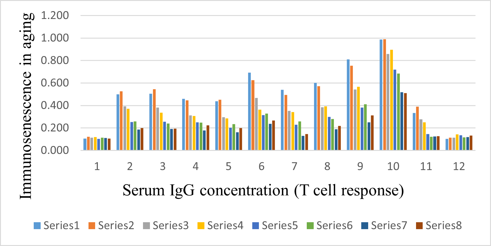
Figure 1: BAR graph from ELISA PLATE
(Source: Created by the learner)
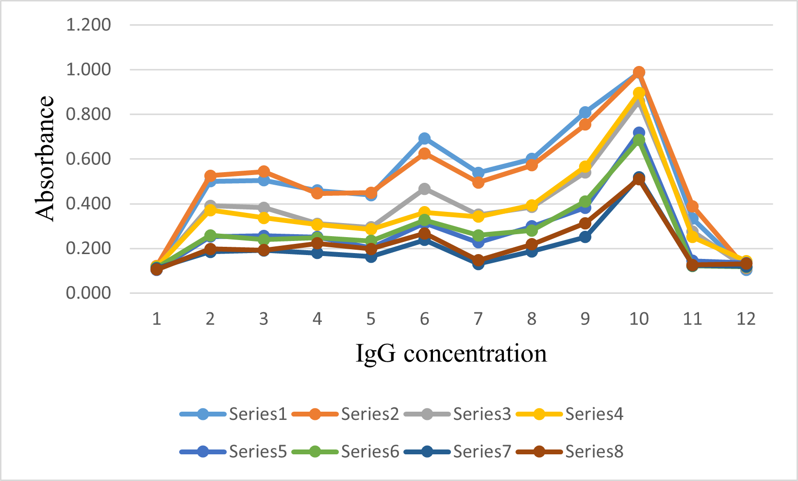
Figure 2: Standard curve from ELISA PLATE
(Source: Created by the learner)
The endpoint titration of ELISA is for determining both the level and response of antibodies. T lymphocyte is responsible for regulating immunity response as well as carrying out various effector functions. T cell that explicit CD8 molecules hold the potentiality to lyse the target cell. It is too evidenced that the cytotoxic T lymphocytes (CTL) act as a major effector for tumor rejection (Song et al. 2018). It is examined that for both humans and mice, T cells are vital to generating an effective as well as long-lasting “cytotoxic CD8 T-cell response”. moreover, the subset of T lymphocytes is taking part in regulating immune response through cytokine secretion as well as antigen-presenting cell activation. T cell destruction includes unresponsiveness of T lymphocyte and lack of antigen which result from ineffective priming along with tumor growth (Ventura MT et al. 2017). Molecular recognition of tumor epitope used to identify by T lymphocytes through a novel immunotherapeutic procedure for expanding tumor-specific T cells. It is also examined that T cell response is cell-mediated as well as a humoral immune response which may control disease enhancement. Cytokines have been tested from supernatants T cell lines through cytomix assay and screening assay (Egorov et al. 2018). Antigen-specific T cell lines have been generated here for getting immunodominant epitope in the ELISA plate. High-throughput epitope mapping has been applied for simultaneous manipulation of the individual immunodominant plates.
Discussion and conclusion
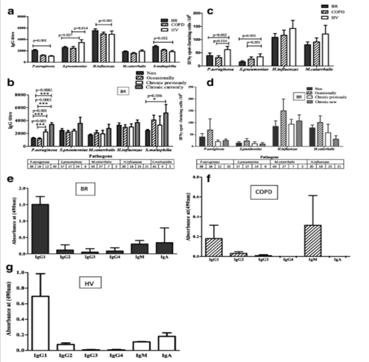
Depending on the antigens in the ELISA plate, the incubation period, the concentration of the used buffer, and the experimental end time vary. The linear range of ELISA (serial dilution) can give superior assay results in comparison with single dilution measurements (Koutsakos et al. 2021). Moreover, the high ELISA assay signals require different assay buffers, operation, titration, specificity, and three independent laboratories for setting up ELISA ADA assay. Here, the preliminary ELISA result has demonstrated detectable antibodies in this observed assay. The comparison of ELISA and cellular assay formal cannot be performed with identical samples. Based on the data from T cell response concerning antibacterial antibody, T cell response is lower in older adults suffering from BR or COPD in comparison with elder people having HIV (Refer to Appendix 3). It is observed that there are no such differences in material method and physical analysis of ELISA in different research papers.
The quantity and quality of T cell responses change overages with resulting in changes in the effectiveness of immune responses. The ineluctable consequence of various scenarios may reduce the potentiality of elder people in responding towards novel antigens or vaccines which enhance susceptibility to infectious disease (Mogilenko et al 2021). It is evidenced that the use of growth factors for T cell function towards checkpoint inhibitor can improve immunity response elderly and proper combination of receptor agonists are enable to increase the effectiveness of vaccination too among older adults. Several studies have shown that antigens play a significant role in shaping the compartment of T cells during aging. The assay-mark of immunosenescence involves reduce potentiality to respond against new antigens, an assemblage of memory T cells, and surviving level of second-rate inflammation (inflame-aging) as well. Mechanistically, immunosenescence can be illustrated partially through cellular and organismal senescence (Jaat FG et al. 2018). it is also identified that T cells are generous among youth but gradually used up by influence to microorganisms throughout life and its number start decreasing within primary lymphoid organs. Apart from that, after the age of 55, the production of T cells becomes 10% lesser than the peak value and that also impacts on immunological space of older people. Age-linked decrease of T cell responses and diminution T cell repertoire has been observed in various human populations. The long-term microbial burden may induce advanced enablement of macrophages, therefore developing low-grade inflammation or inflamm-aging (authentication of immunosenescence).
As per the autoimmune theory, through ages, the immune system going to lose its effectiveness and elder people suffer from widespread autoimmune dysfunction. Age-linked proceeding that results in autoimmune disease includes an accumulation of senescent cells and heterogeneous within target tissue as well as the immune system (Mogilenko et al 2021). On the other hand, based on immunodeficiency theory, the immune system towards novel antigens used to rely on T cell availability as well as age-associated thymic involution. Moreover, the age-linked decrease of thymic output signal effect on T cell reservoir and make it prone to both non-infectious and infectious disease. In this paper, though the contribution of immunosenescence in aging concerning T cell responses has been explained still how these immune response changes are not illustrated clearly. Besides, cell cytometry has been discussed here but the signaling pathway of T cell responses among the elder has not been emphasized. for improving the research, the ELISA kit instruction must be checked, and all recommended washing protocols ought to be followed. ELISA plate should be incubated at the proper temperature and at shaking speed. Apart from that, sample number must be determined and reagent availability should be verified before the assay. Most importantly, during the assay, reagent, buffer concentration, and lot number must be recorded with accurate deviations. The environmental condition must be recorded as it can impact ELISA performance and proper pipetting technique ought to be followed as well (Oh SJ et al. 2019).
Experiments and analysis can be upgraded for T cell secretes cytokines through ELISA kits and quantification of sensitive protein (Refer to Appendix 4). Flow cytometry is another powerful tool that can provide insight into the immunophenotyping of T cells. Flow cytometry and ELISA are significant in quantitative analysis of T cell responses and expression of its surface antigens. Apart from that flow cytometry is useful in detecting intracellular markers and facilitating intracellular penetration of target antibodies. Understanding immune regulation and age-related disorders are vital for identifying effective strategies for immune rejuvenation and T cell responses.
Reference
Bektas A, Schurman SH, Sen R, Ferrucci L. Human T cell immunosenescence and inflammation in aging. Journal of leukocyte biology. 2017 Oct;102(4):977-88. Available from https://pubmed.ncbi.nlm.nih.gov/28733462/
Egorov ES, Kasatskaya SA, Zubov VN, Izraelson M, Nakonechnaya TO, Staroverov DB, Angius A, Cucca F, Mamedov IZ, Rosati E, Franke A. The changing landscape of naive T cell receptor repertoire with human aging. Frontiers in Immunology. 2018 Jul 24;9:1618. Available from https://internal-journal.frontiersin.org/articles/10.3389/fimmu.2018.01618/full
Ewer KJ, Barrett JR, Belij-Rammerstorfer S, Sharpe H, Makinson R, Morter R, Flaxman A, Wright D, Bellamy D, Bittaye M, Dold C. T cell and antibody responses induced by a single dose of ChAdOx1 nCoV-19 (AZD1222) vaccine in a phase 1/2 clinical trial. Nature medicine. 2021 Feb;27(2):270-8. Available from https://www.nature.com/articles/s41591-020-01194-5?
Gurevich V, Kotharu P, McCann K, Bertolini J. Improvement of ELISA procedures through simultaneous addition of antigen and detection antibody and elimination of washing steps. Journal of Immunoassay and Immunochemistry. 2017 Sep 3;38(5):494-504. Available from https://www.tandfonline.com/doi/abs/10.1080/15321819.2017.1331171
Habel JR, Nguyen TH, Van De Sandt CE, Juno JA, Chaurasia P, Wragg K, Koutsakos M, Hensen L, Jia X, Chua B, Zhang W. Suboptimal SARS-CoV-2? specific CD8+ T cell response associated with the prominent HLA-A* 02: 01 phenotype. Proceedings of the National Academy of Sciences. 2020 Sep 29;117(39):24384-91. Available from https://www.pnas.org/content/117/39/24384.short
Jaat FG, Hasan SF, Perry A, Cookson S, Murali S, Perry JD, Lanyon CV, De Soyza A, Todryk SM. Anti-bacterial antibody and T cell responses in bronchiectasis are differentially associated with lung colonization and disease. Respiratory research. 2018 Dec;19(1):1-2. Available from https://link.springer.com/article/10.1186/s12931-018-0811-2
Koutsakos M, Sekiya T, Chua BY, Nguyen TH, Wheatley AK, Juno JA, Ohno M, Nomura N, Ohara Y, Nishimura T, Endo M. Immune profiling of influenza?specific B?and T?cell responses in macaques using flow cytometry?based assays. Immunology assignment Immunology and Cell Biology. 2021 Jan;99(1):97-106. Available from https://asi.onlinelibrary.wiley.com/doi/abs/10.1111/imcb.12383
Mogilenko DA, Shpynov O, Andhey PS, Arthur L, Swain A, Esaulova E, Brioschi S, Shchukina I, Kerndl M, Bambouskova M, Yao Z. Comprehensive profiling of an aging immune system reveals clonal GZMK+ CD8+ T cells as conserved hallmark of inflammaging. Immunity. 2021 Jan 12;54(1):99-115. Available from https://www.sciencedirect.com/science/article/pii/S1074761320304921
Oh SJ, Lee JK, Shin OS. Aging and the immune system: the impact of immunosenescence on viral infection, immunity and vaccine immunogenicity. Immune Network. 2019 Dec 23;19(6). Available from https://synapse.koreamed.org/articles/1140048
Song Y, Wang B, Song R, Hao Y, Wang D, Li Y, Jiang Y, Xu L, Ma Y, Zheng H, Kong Y. T?cell Immunoglobulin and ITIM Domain Contributes to CD 8+ T?cell Immunosenescence. Aging cell. 2018 Apr;17(2):e12716. Available from https://onlinelibrary.wiley.com/doi/abs/10.1111/acel.12716
Ventura MT, Casciaro M, Gangemi S, Buquicchio R. Immunosenescence in aging: between immune cells depletion and cytokines up-regulation. Clinical and Molecular Allergy. 2017 Dec;15(1):1-8. Available from https://link.springer.com/article/10.1186/s12948-017-0077-0
Appendices
Appendix 1:Steps of ELISA
Appendix 2: Standard curve
Appendix 3: T cell response and antibacterial antibody
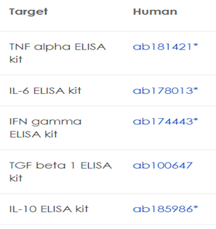
Appendix 4: ELISA kits
